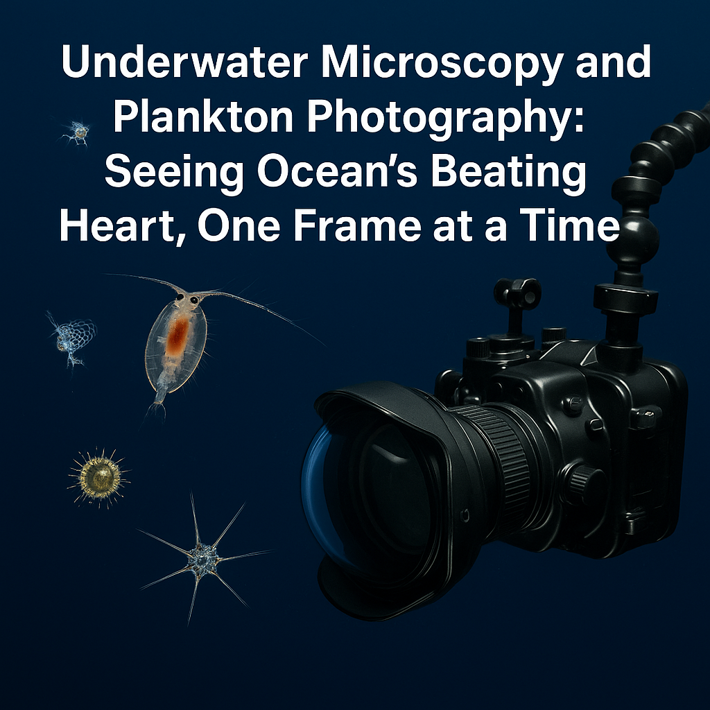
Discover how underwater microscopy and plankton photography are transforming ocean science and maritime operations—from HAB early warnings and aquaculture health checks to education, art, and policy. Explore key tools (IFCB, ISIIS, benthic microscopes, PlanktoScope), best-practice field methods, case studies, FAQs, and a forward look at AI and satellite fusion.
The moment a raindrop turns into a galaxy
Peer through a microscope at a drop of seawater and you’ll watch a living galaxy come into focus: glass-shelled diatoms spinning like satellites, copepods beating invisible oars, larval fish learning to swim, and chains of cyanobacteria painting the water green. For most of us, the ocean is waves and whales. For the ocean itself, life begins with plankton—the drifting plants, animals, and microbes that underpin seafood, blue-carbon cycles, and the health of ports and coasts.
Until recently, studying these tiny engineers meant hauling buckets, preserving samples, and counting cells by eye—slow, biased, and often too late to catch an algal bloom’s first whisper. Today, underwater microscopy and plankton photography let us measure life in situ, in real time, at micrometre scales and over entire coastlines. This article is your field-ready, human-centred guide to the tools, methods, and decisions behind the images: where to start, what to buy or build, how to deploy safely around vessels and aquaculture, and how to use imagery to answer questions that matter—from harmful algal bloom (HAB) early warnings to mariculture QA and citizen science.
Why this topic matters in modern maritime operations
Early warnings that save money—and seasons
Underwater imagers can spot HAB species days to weeks earlier than manual monitoring, letting port health teams, marinas, and farms adjust operations before toxins spike or oxygen crashes. For shellfish growers and finfish farms, an extra week of lead time can mean harvest saved and mortality avoided.
Operational safety, efficiency, and compliance
Plankton structure influences water clarity, biofouling pressure, and intake risks for power plants and desalination near ports. Real-time imaging improves risk assessments for cooling water, SDC intakes, and ballast water treatment performance, supporting compliance checks and maintenance.
Education, outreach, and workforce skills
Nothing hooks the next cohort of mariners and marine techs like live plankton video. Imaging supports STCW-aligned training modules (bridge resource management meets environmental awareness), helping crew understand why ballast and biofouling rules exist and how environmental conditions change voyage-to-voyage.
Climate and ecosystem intelligence
Plankton images tell us who’s actually there—not just “chlorophyll went up.” Species-resolved pictures feed carbon budgets, fisheries models, and restoration metrics (kelp, eelgrass, oysters). This granularity makes your environmental reports and adaptation plans more credible.
Quick stat to ground it: roughly half of Earth’s oxygen comes from oceanic plankton; a single tiny bacterium, Prochlorococcus, may generate up to one-fifth of global oxygen.
Foundations: what we mean by underwater microscopy and plankton photography
-
Underwater microscopy: Optical systems that resolve micrometre-scale details in situ (often diver-operated) on the seabed or in the water column. These can image benthic films, coral polyps, kelp epiphytes, and settling larvae without removing them. A landmark instrument—Scripps’s Benthic Underwater Microscope—achieved near-micrometre resolution directly on the seafloor.
-
In-water plankton imaging: Flow-through or shadowgraph systems that image free-swimming plankton in the water column (e.g., IFCB, ISIIS, Scripps Plankton Cameras), often coupled with sensors (temperature, salinity, oxygen). These platforms generate time series suitable for machine-learning classification and early warnings.
-
On-deck and lab imaging: FlowCam and open-hardware systems like PlanktoScope create high-throughput datasets from samples collected by Niskin bottles, plankton nets, or bucket grabs—perfect for education, QA/QC, and community science.
-
Synoptic context from space: Ocean-color satellites (MODIS, VIIRS, Sentinel-2; now NASA PACE) map bloom extent and type, guiding where to deploy microscopes or nets. Imagery reveals plankton fronts, coccolithophore swirls, and HAB plumes.
Key technologies and developments driving change
Diver-operated benthic microscopes: corals, kelp, and micro-dramas
The Benthic Underwater Microscope (BUM) showed that you can watch coral polyps feed, battle, and bleach at sub-cellular scales in the wild. The latest diver-operated platforms add fluorescence channels to probe photosynthesis and symbiont health in real time—powerful for reef managers and restoration sites.
Where it shines:
-
Coral reef health checks (polyp behaviour, algal competition)
-
Kelp-holdfast fouling and recruitment studies
-
Biofilm succession on eco-moorings and living-shoreline modules
Imaging FlowCytobot (IFCB): automated “cell portraits” in flow
IFCB combines flow cytometry with high-speed imaging, capturing thousands of cell images per hour and measuring fluorescence and light scatter for each captured particle—ideal for species-level HAB surveillance and long time series. Deployed units have delivered early warnings of HABs that manual programs missed.
Where it shines:
-
HAB early warning at shellfish leases and power-plant intakes
-
Long-term ports and estuary observatories
-
Training image libraries for ML classifiers (Random Forests, CNNs)
ISIIS and shadowgraph “wide-angle” imagers
The In-Situ Ichthyoplankton Imaging System (ISIIS) photographs delicate zooplankton (larval fish, gelatinous predators) while profiling through the water column—no nets, no damage. It links imagery to hydrography, revealing where baby fish actually hang out relative to fronts and oxygen minima.
Where it shines:
-
Fisheries recruitment studies
-
Offshore environmental baselines for wind and cable routes
-
Night/day surveys without gear biases
FlowCam and flow-imaging microscopes
Commercial FlowCam platforms produce crisp, annotated imagery of phytoplankton and small zooplankton with automated size/shape metrics—a boon for mariculture QA, regulatory monitoring, and university teaching labs.
Where it shines:
-
Routine farm and hatchery checks
-
HAB species confirmation alongside IFCB alerts
-
Undergraduate labs and outreach
Open-hardware: PlanktoScope for the many, not the few
The PlanktoScope is a low-cost, modular, open-source imaging platform designed for labs, schools, sailboats, and citizen science networks. Peer-reviewed work documents high-throughput quantitative imaging with reconfigurable optics, empowering communities to build comparable datasets without big budgets.
Where it shines:
-
Community monitoring in bays and lagoons
-
Offshore racing and cruising fleets contributing transects
-
Teaching method development (from optics to ML)
Satellites join the party: tasking microscopes from space
New missions like NASA’s PACE sharpen our ability to differentiate phytoplankton groups from orbit. Pairing PACE/VIIRS/Sentinel-2 with in-water imagers creates scalable bloom intel: satellites guide where to sample; microscopes provide species-level truthing for models and regulators.
Practical fieldcraft: from “pretty pictures” to defensible decisions
1) Designing a fit-for-purpose imaging plan
-
Start with a decision, not a device. Are you trying to protect a hatchery intake? Prove a bloom met regulatory thresholds? Teach a class? Your goal sets resolution, depth, power, and storage needs.
-
Layer tools. Use satellites to choose days and locations; tow ISIIS or cast nets for mesoscale context; deploy IFCB for species-resolved time-series; spot-check benthic surfaces with a diver microscope.
-
Mind the workflow. Images are only useful if labelled and searchable. Plan filenames, metadata (GPS, depth, optics, exposure), and storage before leaving the dock.
2) Sampling that respects ships, farms, and MPAs
-
Navigation & safety: Coordinate with harbour masters/Coast Guard for work-area notices if towing imagers or deploying moorings.
-
Aquaculture etiquette: Agree on intake proximity and biosecurity—disinfect gear, especially between bays.
-
Protected areas: Some MPAs restrict permanent mounts; use temporary plates or drop cameras instead.
3) Light, optics, and motion—taming the underwater studio
-
Light matters: In the water column, strobes/LEDs should freeze motion without scaring subjects; in benthic work, cross-polarization reduces glare from biofilms.
-
Motion control: For flow-through systems, maintain laminar flow and consistent velocities to avoid motion blur.
-
Calibration: Photograph stage micrometers and color charts at start/end of each campaign; record dark frames for noise subtraction.
4) Image analysis and the AI layer
-
Classify with care: Even great models drift. Re-train classifiers seasonally, validate on held-out sites, and quantify precision/recall per class (especially for regulated HAB taxa).
-
Trust but verify: Use a human-in-the-loop for rare or high-consequence classes (e.g., Karenia brevis, Alexandrium), and confirm with qPCR/HPLC when stakes are high.
-
Share truth: Publish small, curated image sets with labels to help the community improve models; this pays back when others post theirs.
In-depth analysis: what different platforms see and solve
Benthic microscopes: micro-ecology in real time
On coral reefs and kelp holdfasts, life’s critical events—symbiont bleaching, calcifier dissolution, turf-algae overgrowth—happen at micron scales. Diver-operated microscopes let managers watch processes instead of inferring them. These tools document before/after effects of construction, dredging windows, and eco-mooring trials with persuasive visuals to share with stakeholders.
IFCB: turning harbours into living time-series
IFCBs moored at harbour mouths capture continuous portraits of phytoplankton and small protists. In multiple deployments, IFCB streams provided HAB early warnings that manual counts would have missed—buying precious days for closures and farm responses. A submersible unit typically images up to ~10 cells per second and can run for months.
For maritime users: pair IFCB data with intake alarms (chlorination/UV set-points), notification protocols, and contingency harvest plans.
ISIIS: following larvae through fronts and filaments
Plankton ride filaments, eddies, and density steps. ISIIS reveals how larval fish congregate along thermohaline features, sometimes metres wide—behaviour missed by bottle casts. If you’re planning cable routes, offshore aquaculture, or wind-farm surveys, ISIIS-style transects give baseline biodiversity without net damage, and link it to the physical setting for predictive models.
FlowCam & PlanktoScope: making monitoring doable
A small lab can screen dozens of samples per day with FlowCam, flag suspect frames, and then confirm with molecular assays. If budgets are tight, an open-hardware PlanktoScope plus a robust SOP produces comparable size/shape metrics and photographs that transform teaching and local decision-making.
Challenges and solutions (the honest part)
“We’re drowning in data, starving for answers.”
Solution: Make the question the unit of work, not the image. Build dashboards where each panel answers one operational query (e.g., “Is Alexandrium trending up?”). Automate ingestion → classification → QA flags → alert thresholds. Archive raw imagery; report only the metrics that drive decisions.
“HAB alerts came too late.”
Solution: Combine IFCB or FlowCam time-series with weekly eDNA at sentinel sites. Agree a rapid-response ladder: when classifier confidence crosses a predefined threshold for a priority taxon, trigger confirmatory lab tests and pre-closure notices. Keep an “if this, then that” SOP on the wall.
“Images are pretty, but auditors want numbers.”
Solution: Use standard morphometrics (equivalent spherical diameter, biovolume estimates) and link to regulatory thresholds. Where possible, coordinate with national authority methods (shellfish sanitation programs, water boards). Ensure every plot has n, date/time, device, optics, threshold.
“Cameras drift, optics fog, pumps clog.”
Solution: Treat imagers like engines: daily checks, preventive maintenance, spare seals and desiccant, and flow-rate calibrations logged in a simple CMMS sheet. Keep a dummy dataset for software testing when hardware is down.
“Stakeholders don’t trust black-box AI.”
Solution: Explain model limits in plain language. Show confusion matrices and a gallery of near-misses so decision-makers see strengths and weaknesses. Pair AI calls with human QA for high-consequence outcomes.
Case studies / real-world applications
1) HAB early warning for a shellfish bay
Context: A shellfish cooperative lost two peak harvests to late HAB closures.
Action: The co-op installed an IFCB near the main inlet, added weekly qPCR confirmation for priority toxins, and trained staff on classifier QA.
Outcome: Two seasons later, the team issued voluntary early harvest twice based on trend flags; neither event progressed to closure levels. The co-op saved an estimated €480k in avoided loss and overtime.
2) Port environmental team ties satellites to microscopes
Context: A busy estuary faced recurrent blooms that complicated cooling-water intake operations.
Action: The port authority subscribed to Sentinel-2 coastal scenes and PACE previews; bloom anomalies triggered FlowCam triage and roving IFCB deployments at the intake and shipping channel.
Outcome: Over one summer, the port reduced unscheduled intake maintenance by syncing chlorination cycles to plankton composition, not just turbidity.
3) Coral-reef reserve justifies dredging windows
Context: A small harbour expansion needed to prove that turbidity controls protected nearby reefs.
Action: A diver-operated microscope documented polyp behaviour and biofilm shifts before, during, and after activity; work paused when micro-signs of stress appeared.
Outcome: The reserve authority approved a narrower dredging window the next season with stricter triggers, and public trust rose thanks to transparent imagery.
4) Community science by sail
Context: A coastal NGO wanted to extend monitoring beyond a single bay.
Action: Ten citizen crews built PlanktoScopes and followed a simple SOP: fixed tow speed, depth bucket, standard volume, same lighting, and an upload app.
Outcome: Over one year they produced the region’s first seasonal plankton atlas, informing eelgrass and oyster restoration siting.
How to choose your first (or next) system
If you run a mariculture site:
-
Start with FlowCam or PlanktoScope for lab screening; add IFCB if HAB pressure is high and you need continuous data.
-
Budget for qPCR kits on priority taxa and a simple classifier tuned to your bay.
If you’re a port or power plant EHS lead:
-
Subscribe to satellite ocean-color feeds and turbidity/fluorescence sensors at intakes.
-
Pilot in-water cleaning with capture for biofouling on service craft; image scrapings to build a local fouling library that informs coating choices and intervals.
If you’re an educator or outreach lead:
-
Build a PlanktoScope, pair with a DSLR on a stereoscope, and design a “plankton to policy” lab: show images → connect to ballast/biofouling rules → discuss bloom risk and seafood safety.
-
Invite mariners and aquaculture techs—cross-pollination builds understanding.
If you’re a reef or restoration manager:
-
Borrow or partner on a benthic microscope for seasonal campaigns; link images to temperature and pH loggers.
-
Photograph eco-moorings, living-shoreline modules, and kelp recruitment plates to show tangible outcomes.
Future outlook: where this is all going
-
AI that explains itself: Classifiers will deliver per-image rationales (“these diatom ribs made me call Pseudo-nitzschia”), improving auditability.
-
Port eDNA + image fusion: Monthly eDNA screens will sit alongside IFCB/FlowCam time series for invasive and HAB detection, normalised like routine water sampling.
-
PACE-era habitat nowcasts: Satellite hyperspectral fingerprints will flag bloom types; ports and farms will task mobile imagers accordingly.
-
Open, comparable datasets: Standard image metadata and shared taxon libraries will make cross-port comparisons meaningful, accelerating learning.
-
Diver microscopes with structured light: Next-gen benthic systems will map 3D micro-topography alongside fluorescence, linking form and function on reefs and kelp.
Frequently asked questions (FAQ)
Isn’t chlorophyll a enough—why image?
Chl-a says “biomass up/down.” Images say who. For operations and health, composition matters: diatoms vs. dinoflagellates, toxin producers vs. harmless bloomers.
How early can an IFCB warn of a HAB?
In practice, days to weeks—enough to change harvest timing or prep mitigation—because it runs continuously and classifies species, not just pigments.
Do I need a scientist on board to use these tools?
You need good SOPs and at least one trained tech. Partner with a local lab or university to calibrate and QA your first season. The open-hardware path keeps costs down.
Will satellites replace microscopes?
No—they complement. Satellites tell you where and when; microscopes tell you who and how much. Together they make monitoring faster and cheaper.
What about data overload and AI errors?
Start with a short priority list (e.g., six taxa of concern). Track precision/recall; keep a human-in-the-loop for high-stakes calls; and archive raw images for re-analysis as models improve.
Can students or citizen scientists contribute useful data?
Absolutely. With PlanktoScope SOPs and basic QA, communities produce consistent, usable imagery that fills gaps between official stations.
Conclusion: Pictures that make better choices
Underwater microscopy and plankton photography are not just eye-candy. They’re decision tools—for farmers scheduling harvests, captains planning intakes, port teams managing risks, teachers lighting curiosity, and policymakers setting thresholds. When you can see the plankton, you can act sooner, target better, and explain your choices to partners and the public.
Start simple: set your goal, pick one platform, write an SOP, and share your first gallery with the people who depend on a healthy, predictable coast. In a world of changing seas, the most beautiful pictures are the ones that help communities thrive.
References (hyperlinked)
-
NOAA Ocean Service. (2024). How much oxygen comes from the ocean? https://oceanservice.noaa.gov
-
Woods Hole Oceanographic Institution (Anderson/Sosik labs). Imaging FlowCytobot (IFCB) overview & technical background. https://www2.whoi.edu
-
Northeast HAB (WHOI). IFCB field performance and throughput. https://northeasthab.whoi.edu
-
Mullen, A. D., et al. (2016). Underwater microscopy for in situ studies of benthic ecosystems. Nature Communications. https://www.nature.com/articles/ncomms12093
-
Smithsonian Ocean. In-Situ Ichthyoplankton Imaging System (ISIIS). https://ocean.si.edu
-
Oregon State University — Hatfield Marine Science Center. ISIIS overview. https://hmsc.oregonstate.edu
-
Fluid Imaging (FlowCam). Plankton research applications. https://www.fluidimaging.com
-
Pollina, T., et al. (2022). PlanktoScope: Affordable modular quantitative imaging of plankton. Frontiers in Marine Science. https://www.frontiersin.org/journals/marine-science/articles/10.3389/fmars.2022.949428/full
-
NASA Ocean Color. Basics and instruments; PACE program learning pages. https://oceancolor.gsfc.nasa.gov ; https://pace.oceansciences.org
-
Campbell, L., et al. (2013). Continuous automated imaging-in-flow for early warning of HABs (IFCB). (Open-access summary). https://pubmed.ncbi.nlm.nih.gov/23307076/
-
Scripps Institution of Oceanography (News). (2025). Diver-operated microscope advances coral health imaging. https://scripps.ucsd.edu
-
TIME Magazine. (2024). Why NASA’s PACE mission is studying phytoplankton and aerosols. https://time.com


Thank you